This review is intended to provide an overview of Pocket Brain, the new interactive neuroanatomy app by eMedia and compare its features to other other iOS based neuroanatomy apps. In short, Pocket Brain is the most comprehensive, interactive, and functional neuroanatomy app out there. It is worth the investment of any student studying neuroanatomy.
Neuroanatomy as a Class
To be good at neuroanatomy a student needs decent visual-spatial reasoning skills. Some are gifted in this regard others are not. I took clinical neuroanatomy before I had an iPad. The amount of time I dedicated (or wasted) flipping through my neuroanatomy atlas to remind myself of the location and function of each particular structure was tremendous, and I’m certain that I was not alone in this regard. Post-iPad purchase, when it came time to study for USMLE Step 1, I used nearly every neuroanatomy app out there including: Brain Tutor HD, 3D Nervous System, FINR Brain, and 3D Brain. Of these apps, the last two: FINR Brain, and 3D brain were my “go-to” apps. While they didn’t have everything I needed, each app served its purpose and got me one step closer understanding the important spatial and functional relationships needed to master neuroanatomy.
Pocket Brain by eMedia
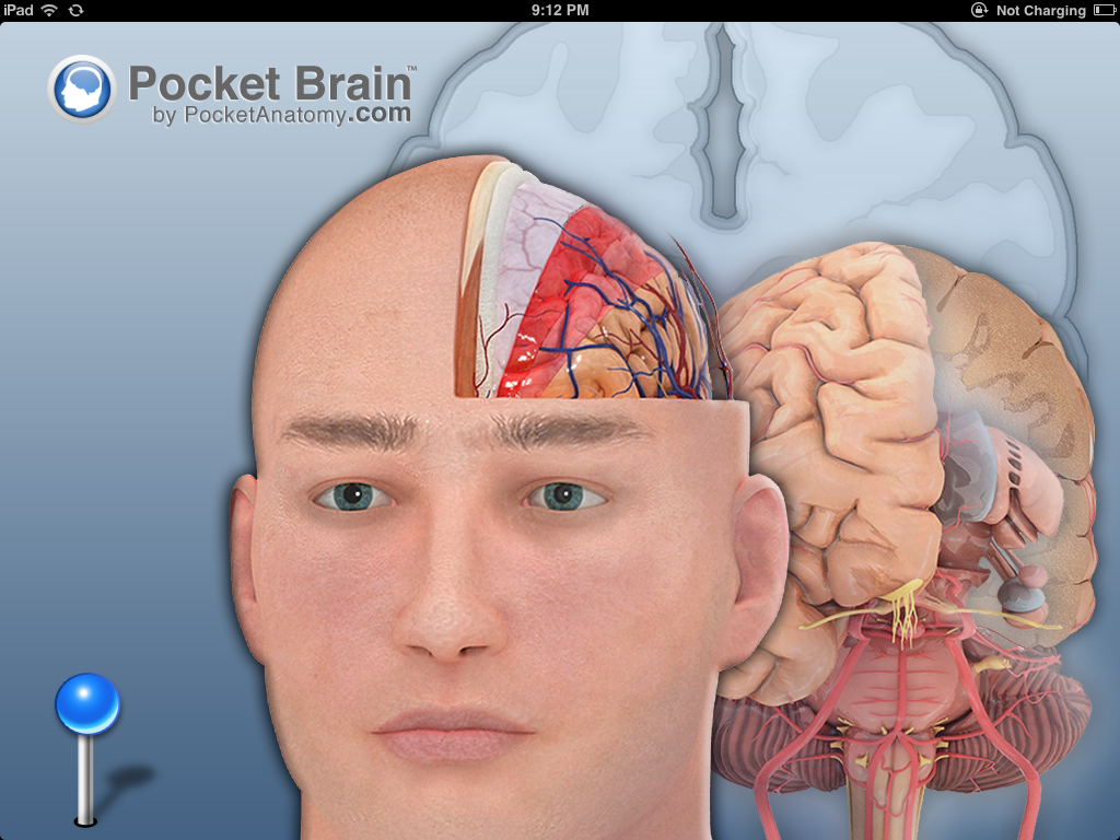
Pocket Brain takes interactive neuroanatomy to a whole new level. As a disclaimer, I fell in love with eMedia’s apps when they created “Pocket Body” which is their interactive anatomy app focused on the musculoskeletal system. They have successfully used a similar feature set with Pocket Brain.
4 Components of the Pocket Brain
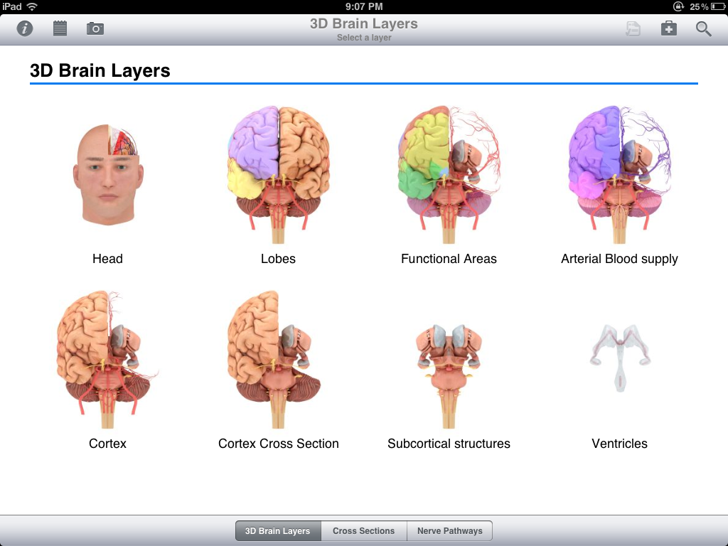
There are 4 main components to Pocket Brain: (1) 3D Brain Layers (which is best part of the app) (2) Cross Sections (3) Nerve Pathways (4) Quizzes and clinical cases.
(1) 3D Brain Layers
The “3D Brain Layers” part of the app follows in Pocket Body’s tradition by allowing the user to add and remove “anatomical layers” by sliding your finger or pressing a button on the screen. This “layer slider” is rather intuitive and easy to use.
If your unfamiliar with the “Layer Slider” and not sure how it works, just imagine the brain is an onion. Every time you use the layer slider to remove a layer this would be analogous to peeling an onion from left to right while keeping the core intact. Eventually you remove the last layer and you are left with the core of the onion. In Pocket Brain this last layer would be the ventricles of the brain. What is nice about using this app and the iPad compared to a text book is that as you work through each layer you can rotate the anatomical structures to get unique view points about 2 different axes.
Each layer is pre-defined and serves a purpose. Note: In the past (with Pocket Body) some of the layers seemed somewhat arbitrarily defined, however with Pocket Brain, eMedia has done a better job at giving each layer specific utility.
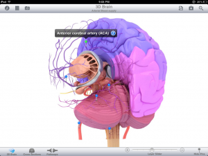
 For example there is a layer titled “Arterial blood Supply.” In this layer, each part of brain is color coded to represent its major arterial supply. So areas of the brain supplied by the anterior cerebral artery (ACA) are shaded blue, while the areas supplied by the middle cerebral artery (MCA) are shaded in purple. What I found very beneficial with Pocket Brain is that you don’t have obtrusive text all over the page.
Instead of text, each label is replaced by a pin which the user taps on into to see the label and additional information about the anatomical structure. This additional info is organized 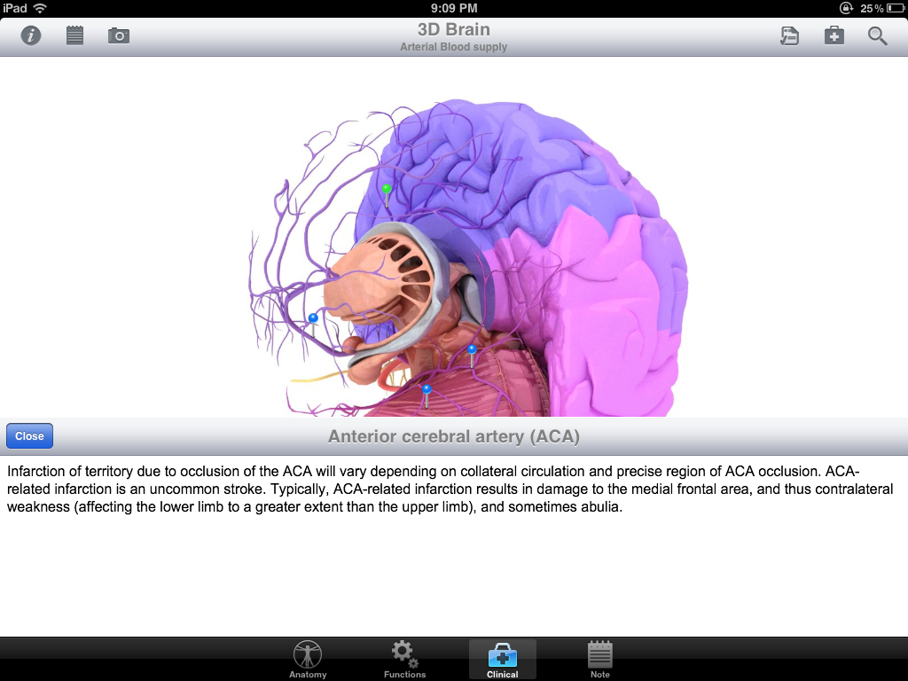 into 4 meaningful sections with subtopics titled, (1) Anatomy, (2) Functions (3) Clinical, (4) Notes. This is shown on the left. The titles are descriptive of the information. Anatomy – talks about anatomical relationships, Functions – talks about the physiologic functions, Clinical – adds some sort of clinical correlate, Notes – allows the user to add and store their own notes tagged to each structure.
This organization of information is really is where Pocket Brain excels and adds value to the student. This is what is unique about Pocket Brain when compared to a typical atlas and most other neuroanatomy apps.
(2) Cross Sections
Brain cross sections are probably some of the hardest things to learn, but arguably the most clinically relevant because this is what you’ll be seeing in any sort of diagnostic imaging.
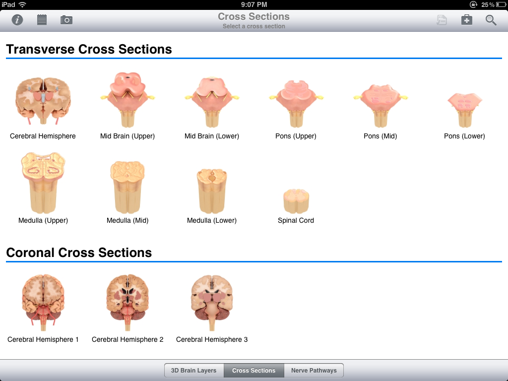
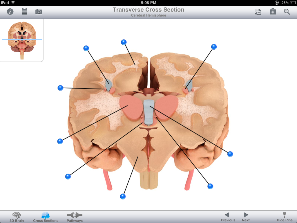 Included are 10 transverse cross sections and 3 coronal cross sections (as shown at the top of this post). In terms of details, it seems like they tried to strike a balance and not overwhelm the user. If you compare it to the utility and majestic-ness of the 3D Brain layers you are likely to be underwhelmed. It is a good start, but what would make this part of the app more useful is if they added some diagnostic imaging here (MRI, CT scan) so that you see both at the same time, possibly with the outline of the drawing overlapping the MRI (Imagine two overhead transparency slides stacked on top of one another. Boston University has a great histology website which has a similar feature.)
When it comes to brain cross sections, most neuroanatomy apps lack this completely. Brain Tutor HD does a decent job by having scrollable cross section, however the user interface is poor, and it is too cumbersome to use. I’m happy see Pocket Brain include cross-sections, and I’m confident it will be developed further in future editions.
(3) Nerve Pathways:
This is a high-yield area and an important component of neuroanatomy. It is a shame that it has been neglected in the other apps until now and again I’m happy to see it in Pocket Body. The target audience here seems to be for the novice learner. The app shows the progression of particular sensations from body to cortex. Specifically, you see a highlighted arrow go from the body, through the spinal cord, through the brain stem, and eventually to the cortex. Pocket Brain covers the major 4 tracts: spinothalamic, 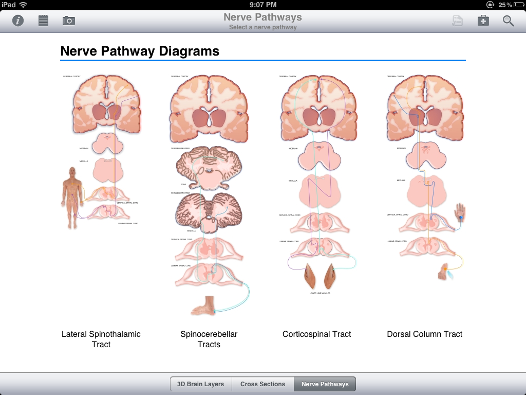 spinocerebellar, corticospinal, and dorsal column. For the USMLE you should be able to analyze an image of a damaged spinal cord and determine what the clinical ramifications may be. You should also be able to identify which parts of the spinal cord would be affected in ALS, multiple sclerosis, or tabes dorsalis. Pocket is missing some of this core information, and thus there is room for improvement. In any case, this is a needed step in the right direction.
(4) Quizzing and Clinical Cases:
Like Pocket Body, there are 3 different types of quizzes which allow the user to test their knowledge (A) Locate an anatomical structure, (B) Flash Card quiz (C) Multiple Choice quiz.
Unique to Pocket Brain, compared to its predecessor Pocket Body, is a section labeled Clinical Cases. Currently, there are only a handful of clinical cases (4 neuromuscular and 4 stroke). Again this is something that I am very happy to see. This is a needed addition to all anatomy apps as clinical problem solving skills is the crux of what most healthcare professionals need to know. As any clinician or seasoned educator will tell you, “What is the point of having knowledge if you cannot put the facts together (inductive reasoning) to solve a problem?” I hope to see this develop in the future, and for you go-getters there is even “suggest a clinical case by email” button to help eMedia improve in this regard. Kudos to Pocket Brain for this.
In comparison to other apps, the FINR app has 9 clinical injuries with well-designed interactive pictures showing where and how the brain is injured. I like this a lot, however there are only limited number of injuries depicted. However with FINR there are no quizzes and the injuries are not presented as a case, but instead the info is presented as a description. Thus FINR may have more value to the doctor describing one of these 9 pre-determined clinical diseases to a patient, whereas Pocket Brain has more value to a student trying to attach clinical symptoms to disease.
Conclusion:
To conclude, the best part of this app is the 3D Brain Layers. It leverages the experience and functionality of the award-winning Pocket Body App and applies it to Pocket Brain. For this reason alone, I believe this app is worth the money.
The $20 price tag for the app makes Pocket Brain more expensive than FINR and 3D Brain tutor. As a student, I understand that it might seem like a large investment, however to put it in context, it is about half the cost of a typical neuroanatomy atlas. Using Pocket Brain while studying neuroanatomy will save you time, and a question you need to ask yourself “How much am I willing to spend to save time and avoid frustration?” The decision is ultimately yours.
Pocket Brain does significantly outshine all of the other neuroanatomy apps in nearly every category including topic coverage, usability, interface, organization, and functionality.
With 3D brain layers, high-yield cross sections, nerve pathways, and quizzing/clinical cases it has most of your bases covered. I give this app, in version 1.0, four and a half stars. While there is room for improvement, the app is currently the best out there and they have laid the foundation to make it better. The “Pocket” series has clearly established itself as a big player in the iPad Anatomy App market, and I look forward to seeing what they come up with next.
Areo Saffarzadeh
Medical Student, Year 3
UC Irvine School of Medicine